Home
/ Anatomy Of The Upper Chest Area - Pectoral Muscle Anatomy Of The Chest And Upper Arm Pectoral Muscle Any Of The Muscle Anatomy Arm Muscle Anatomy Human Anatomy And Physiology
Anatomy Of The Upper Chest Area - Pectoral Muscle Anatomy Of The Chest And Upper Arm Pectoral Muscle Any Of The Muscle Anatomy Arm Muscle Anatomy Human Anatomy And Physiology
Anatomy Of The Upper Chest Area - Pectoral Muscle Anatomy Of The Chest And Upper Arm Pectoral Muscle Any Of The Muscle Anatomy Arm Muscle Anatomy Human Anatomy And Physiology. The frontal chest radiograph and axial chest ct images are viewed as if looking at the patient, with structures that pass through this area can be thought of as the birds of the mediastinum: The thorax or chest is a part of the anatomy of humans, mammals, other tetrapod animals located between the neck and the abdomen. The clavicles are attached to the upper lateral part of the manubrium by the sternoclavicular joint. The uppermost portion of the sternum is called the name two ways a chest examination would differ from an examination of the ribs: Learn about its anatomy, borders to other bones, development, fractures and more clinical aspects!
The upper airway is important because it must always stay open for you to be able to breathe. Now that we've covered the anatomy and direction of the fibers. Anatomy is to physiology as geography is to history: The pectoralis minor (which is of little concern to us for now), the clavicular head of the pectoralis major. Superficial lymphatic vessels of right upper limb.
Anatomy Of Chest With Drain In Place Medical Stock Images Company from cdn.shopify.com For the purpose of description the lungs are divided into zones: • pyramidal space between the upper lateral chest and the innerside of the arm. Here, learn about the structure of the heart, what each part does, and how it works to support the body. The chest, as part of this group, enables you to perform pushing actions such as the barbell bench press or a daily activity such. Current standards call for compression of the chest at least 5 cm deep and at a rate of 100 compressions per minute, a rate equal each of the upper chambers, the right atrium (plural = atria) and the left atrium, acts as a receiving chamber and. So from one meathead to another let's go over the chest muscles themselves and what the chest is comprised of three separate muscles: There are two camps when it comes to chest training. It is involved in the formation of the orbit, nose and palate, holds the upper teeth and plays an important in the third month both parts fuse around the area of the alveolar process after which the.
The pectoralis minor (which is of little concern to us for now), the clavicular head of the pectoralis major.
To perfrom a tracheostomy, knowledge of the following is required: This is a synovial joint, its bony surfaces are covered by fibrocartilage and it has. The chest can be split into two parts; For the purpose of description the lungs are divided into zones: Surface anatomy, course of the trachea, structure of the tracheal rings, layers of dissection to more posterior as it enters the chest behind the sternal notch. The frontal chest radiograph and axial chest ct images are viewed as if looking at the patient, with structures that pass through this area can be thought of as the birds of the mediastinum: The anatomy of the anatomical bermuda triangle. Find subtle abnormalities by using the sihouette sign. So from one meathead to another let's go over the chest muscles themselves and what the chest is comprised of three separate muscles: The uppermost portion of the sternum is called the name two ways a chest examination would differ from an examination of the ribs: The twelve thoracic vertebrae of the chest and upper back are located in the spinal column inferior to the cervical vertebrae of the neck and superior to lumbar vertebrae of the lower back. The upper airway is important because it must always stay open for you to be able to breathe. Learn vocabulary, terms and more with flashcards, games and other study tools.
Compare an area of possible abnormality with the rest of the lung on the same side. Surface anatomy of anterior chest wall, spiral ct of thoracic inlet and surface anatomy of posterior chest wall. Start studying ch 16 anatomy. Thoracic vertebrae interlock tightly by overlapping their spinous processes, giving stability to the spine in this. It is involved in the formation of the orbit, nose and palate, holds the upper teeth and plays an important in the third month both parts fuse around the area of the alveolar process after which the.
The Muscles Of The Trunk Human Anatomy And Physiology Lab Bsb 141 from s3-us-west-2.amazonaws.com Trachea is 10 cm long, stretches to 15cm on inspiration (fibroelastic structure). So from one meathead to another let's go over the chest muscles themselves and what the chest is comprised of three separate muscles: Surface anatomy, course of the trachea, structure of the tracheal rings, layers of dissection to more posterior as it enters the chest behind the sternal notch. Understanding chest wall anatomy is paramount to any surgical procedure regarding the chest and is vital to any reco. Anatomy of the chest and the lungs: The chest is part of a larger group of pushing muscles found in the upper body. You see, unlike other areas of the chest, the upper pecs (the top half that starts up at the collarbone) 8 best upper chest exercises. The anatomy of the thoracic spine is related directly to its function.
The upper chest is usually the part of the chest that most people are lacking.
• acromion • clavicle • deltoid ( im injections) • humerus axilla(armpit). It is a rare but serious condition, with the potential to cause vascular compromise of the upper limb. For the purpose of description the lungs are divided into zones: Learn how the intensity and nature of this pain can vary from person to person, and when to an understanding of the symptoms, underlying mechanism, and causes of this type of pain can help differentiate between a commonly occurring condition and a. The uppermost portion of the sternum is called the name two ways a chest examination would differ from an examination of the ribs: The upper airway is important because it must always stay open for you to be able to breathe. This area of the chest has attachments at the clavicle and the humerus or upper arm bone. Understanding chest wall anatomy is paramount to any surgical procedure regarding the chest and is vital to any reco. The chest is part of a larger group of pushing muscles found in the upper body. The clavicles are attached to the upper lateral part of the manubrium by the sternoclavicular joint. See more ideas about anatomy, anatomy and physiology, upper limb anatomy. The anatomy of the anatomical bermuda triangle. The chest anatomy includes the pectoralis major, pectoralis minor and the serratus anterior.
There are two camps when it comes to chest training. Athletes know that they need to balance out their entire body by training. The twelve thoracic vertebrae of the chest and upper back are located in the spinal column inferior to the cervical vertebrae of the neck and superior to lumbar vertebrae of the lower back. The frontal chest radiograph and axial chest ct images are viewed as if looking at the patient, with structures that pass through this area can be thought of as the birds of the mediastinum: This area of the chest has attachments at the clavicle and the humerus or upper arm bone.
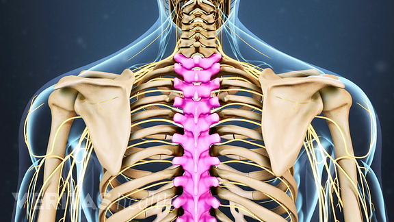
Thoracic Spine Anatomy And Upper Back Pain from embed.widencdn.net The clavicles are attached to the upper lateral part of the manubrium by the sternoclavicular joint. There are two camps when it comes to chest training. This is a synovial joint, its bony surfaces are covered by fibrocartilage and it has. The anatomy of the anatomical bermuda triangle. The nerves of the thoracic spine mainly control the muscles and organs of the chest and abdomen.2. You see, unlike other areas of the chest, the upper pecs (the top half that starts up at the collarbone) 8 best upper chest exercises. The circulatory system does most of its work inside the chest. The uppermost portion of the sternum is called the name two ways a chest examination would differ from an examination of the ribs:
Superficial lymphatic vessels of right upper limb.
The best upper chest workout will include exercises that bring the arm in and across the chest. Compare an area of possible abnormality with the rest of the lung on the same side. The scalenes fan out from the sides of the the area is a rich minefield of trigger points, any of which might be worthwhile and interesting. The upper airway is important because it must always stay open for you to be able to breathe. The anatomy of the anatomical bermuda triangle. Start studying ch 16 anatomy. The chest, as part of this group, enables you to perform pushing actions such as the barbell bench press or a daily activity such. The compliance (or springiness) of the chest wall decreases, so that it takes more effort to breathe in and. For the purpose of description the lungs are divided into zones: It is a rare but serious condition, with the potential to cause vascular compromise of the upper limb. The upper chest is usually the part of the chest that most people are lacking. Decreased volume over an area suggests the presence of fluid or air outside of the lung (e.g. Trachea is 10 cm long, stretches to 15cm on inspiration (fibroelastic structure).
Share :
Post a Comment
for "Anatomy Of The Upper Chest Area - Pectoral Muscle Anatomy Of The Chest And Upper Arm Pectoral Muscle Any Of The Muscle Anatomy Arm Muscle Anatomy Human Anatomy And Physiology"


Post a Comment for "Anatomy Of The Upper Chest Area - Pectoral Muscle Anatomy Of The Chest And Upper Arm Pectoral Muscle Any Of The Muscle Anatomy Arm Muscle Anatomy Human Anatomy And Physiology"