Chest Muscles Diagram Labeled : Upper Limb Labeling Worksheet Printable Worksheets And Activities For Teachers Parents Tutors And Homeschool Families. View the muscles of the upper and lower extremity in the diagrams below. In fact, when you work. Muscle diagrams are a great way to get an overview of all of the muscles within a body region. To get started, choose a muscle group either on the muscle chart. Muscles that act on the chest.
The dominant muscle in the upper chest is the pectoralis major. Chest muscles, chest muscle diagram. Muscles that act on the chest. It's pointing to a lower spot of the rectus femoris. Studying these is an ideal first step before moving labeled diagram.
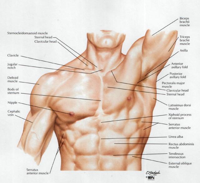
Club 100 Trainer And General Manager Eufay Wood Of High Octane Training With Club 100 Member Mike Holcomb Demonstrate A Few Training Techniques In A Video Series We Are Doing This Month from tahoeclub100.com Chest muscles, chest muscle diagram. The bones shown in the chest and hip region in the labeled human skeleton diagram are the ribs, vertebrae, pelvis, os coxae, sacrum and coccyx. Flexion, extension and abduction of the shoulder. The chest anatomy includes the pectoralis major, pectoralis minor and the serratus anterior. Muscle anatomy types of movement all muscles exert their force by pulling between at least two points of attachment. The movement that results from contraction is called the action of the muscle. Primarily, there are three chest muscles involved in movement: The muscles of the chest are the following ones.
Now label the diagram in your workbook!
Freetrainers.com has a vast selection of exercises which are used throughout our workout plans. This diagram depicts muscle labeled diagram. It's pointing to a lower spot of the rectus femoris. Your chest muscles are big and can handle more weight, which allows you to burn more calories when you exercise them. Muscles of the chest enable us to lift, extend, and rotate our arms, along with playing a part in the process of respiration. Using the word bank, label the muscles shown in the front view on this free worksheet. Download these free printables to teach your kids about the workin. This is one of the internal rotator muscles that attach the humerus and internally rotate the arm. Use the location, shape and surrounding structures to. Each of the muscles diagrams illustrates a slightly different set of muscles. Find stockbilleder af male biceps chest muscle chart labeled i hd og millionvis af andre royaltyfri stockbilleder, illustrationer og vektorer i shutterstocks samling. Female chest muscles with labels. The labeled structures are (excluding the correct side):
The two sides connect at the sternum, or breastbone. Anatomynote.com found labelled diagram of the muscles in the human body from plenty of anatomical pictures on the internet. Freetrainers.com has a vast selection of exercises which are used throughout our workout plans. This is one of the internal rotator muscles that attach the humerus and internally rotate the arm. The bones shown in the chest and hip region in the labeled human skeleton diagram are the ribs, vertebrae, pelvis, os coxae, sacrum and coccyx.
Male Muscle Model from classroom.sdmesa.edu Note how the basilar segmental bronchi are oriented from lateral to medial. View the muscles of the upper and lower extremity in the diagrams below. Breast anterior intercostal artery internal thoracic (mammary) artery costal cartilage body of the sternum xiphisternal joint internal thoracic (mammary) from the case: O muscles—sternocleidomastoid, anterior and middle scalene, infrahyoid normal anatomic structures are labeled on posteroanterior (pa) and lateral chest radiographs (figs. Learn about each of these muscles, their locations the chest is part of a larger group of pushing muscles found in the upper body. The movement that results from contraction is called the action of the muscle. Anatomynote.com found labelled diagram of the muscles in the human body from plenty of anatomical pictures on the internet. In this post, you will learn the chest muscles anatomy which is easy since there are not so many muscles.
The two sides connect at the sternum, or breastbone.
Human anatomy diagrams show internal organs, cells, systems, conditions, symptoms and sickness information and/or tips for healthy living. The sartorius is definitely labeled wrong. Use the location, shape and surrounding structures to. Anatomynote.com found labelled diagram of the muscles in the human body from plenty of anatomical pictures on the internet. A chest muscle that pulls the arm in towards the body. The muscles of the chest are the following ones. Primarily, there are three chest muscles involved in movement: Learn vocabulary, terms and more with flashcards, games and other study tools. Flexion, extension and abduction of the shoulder. The movement that results from contraction is called the action of the muscle. Freetrainers.com has a vast selection of exercises which are used throughout our workout plans. This is one of the internal rotator muscles that attach the humerus and internally rotate the arm. Labeled human anatomy diagram of male biceps, arm, and chest muscles frontal anterior view on a white background.
The labeled structures are (excluding the correct side): Learn muscles anatomy and reference. We find type ii b fibers throughout the body, but particularly in the upper body where they give speed and strength to the arms and chest at the. This diagram depicts muscle labeled diagram. The chest muscles are responsible for moving the arms across the body and up and down, as well as other movements like flexion, adduction, and rotation.
Labeled Atlas Of Anatomy Illustrations Of The Dog from www.imaios.com Male muscular system, full anatomical body diagram with muscle scheme, vector illustration educational poster. The chest, as part of this group, enables you to perform pushing. The two sides connect at the sternum, or breastbone. Now label the diagram in your workbook! A chest muscle that pulls the arm in towards the body. Freetrainers.com has a vast selection of exercises which are used throughout our workout plans. Flexion, extension and abduction of the shoulder. Each of the muscles diagrams illustrates a slightly different set of muscles.
Download these free printables to teach your kids about the workin.
Chest muscles, chest muscle diagram. How to build chest muscle fast. Nerve root anatomical structure labeled cross section. It is accomplished primarily by the sternocleidomastoid muscles, with assistance from. Related posts of chest muscles diagram muscle anatomy in leg. Related posts of back chest muscles diagram. The movement that results from contraction is called the action of the muscle. Tusindvis af nye billeder af høj kvalitet tilføjes hver dag. Human muscle system, the muscles of the human body that work the skeletal system, that are under voluntary control, and that are concerned with neck flexion refers to the motion used to touch the chin to the chest. Learn vocabulary, terms and more with flashcards, games and other study tools. Note how the basilar segmental bronchi are oriented from lateral to medial. Studying these is an ideal first step before moving labeled diagram. I often get asked, how can i build thick powerful pecs?
Related posts of back chest muscles diagram chest muscles diagram. The pectoralis major, the pectoralis minor, and the serratus anterior.
Share :
Post a Comment
for "Chest Muscles Diagram Labeled : Upper Limb Labeling Worksheet Printable Worksheets And Activities For Teachers Parents Tutors And Homeschool Families"


Post a Comment for "Chest Muscles Diagram Labeled : Upper Limb Labeling Worksheet Printable Worksheets And Activities For Teachers Parents Tutors And Homeschool Families"